


A vascular puzzle solved
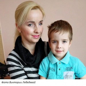 For the smiling Maxim and his mother Elena, Valentine’s Day was truly a holiday: they were discharged from the Children’s Surgery National Applied Research Hospital of the Republic of Belarus. The child was cheerful and happy, as if he had never been ill. Only the bandage and slight nausea reminded of the operation.
The doctors will remember a five-year-old boy for a long time: one meets the patient like this only once in a lifetime. Correction of diaphragmatic hernia and abnormal vascular arterial network in the region of the left lung, esophageal opening of the diaphragm, stomach and liver became a real puzzle. The pediatricians and radiologists started to solve the problem, the surgeons finished.
10 days passed after the surgery, but Elena is still in shock. She describes the history of the disease in detail, recalling everything from the first days of Maxim’s life:
He was born with congenital pneumonia; it was quickly treated. When my son was a year old, I noticed that there was a slight deformation of the chest. The orthopedist advised massage and swimming. The child slowly gained weight, but it seemed that everything was within the norm.
… The boy visited kindergarten, played with his older sister, sometimes was sick, like all children. Last year in autumn, the temperature rose sharply: the diagnosis was pneumonia. The X-ray picture seemed strange to the polyclinic’s radiologist: something incomprehensible was in the left lung and in the field of the diaphragm. The boy was directed on CT. There were two congenital problems – lung sequestration and diaphragmatic hernia.
Pneumonia was quickly cured in one of the city’s children’s hospitals. It was necessary to deal with the diagnosed diseases. Maxim was examined in detail at the Children’s Surgery National Applied Research Hospital.
Alexandr Makhlin, Deputy Clinical Director of the Children’s Surgery National Applied Research Hospital:
THE FIRST such case in the history of pediatric surgery in the republic …
What happened?
Sequestration is a combined malformation of the lungs and vascular system. A part of the body “does not breathe” and is fed by abnormal blood vessels. Turned off from the gas exchange site gives a picture of pneumonia (sometimes – recurrent pneumonia). In the case of Maxim, the pathology affected the lower lobe of the left lung.
The diaphragmatic hernia turned to be the rarest in children – the parasophageal: a part of the stomach presses the heart and left lung. We faced the case like this only a few times in the Children’s Surgery National Applied Research Hospital.
Only surgery can correct the both defects. They are located in one zone, but you can approach the lung only from the side of the thorax, and to cute hernia from the side of the abdominal cavity. How to operate: consistently by two teams or in stages in six months? We stopped on the first option and, as it turned out, not in vain. It took about five hours.
1 st stage. Thoracoscopic intervention on the lungsAlexander Kul, thoracic surgeon, head of the operational unit of the Minsk City Clinical Oncology Center:
Almost all left lung was anatomically in a normal state. Diagnostic angiography revealed an abnormal vessel that receded from the abdominal part of the aorta, reaching the thoracic cavity through the diaphragm. It supplied with blood the area of extrapulmonary sequestration and went further through it into the lower lobe of the lung. Such an abnormal branch led to the fact that part of the oxygen-enriched blood returned to the lungs.
It was necessary to remove the site of extrapulmonary sequestration and block the abnormal blood flow. The vessel was isolated and stitched with the special apparatuses at the diaphragm. Its diameter was 5-6 mm, for a five-year-old child this is the fifth part of the aorta. The difficulty was that the slightest careless movement could lead to massive blood loss, and you would need a thoracotomy … The segregated segment of the lung was removed.
2 nd stage. Correction of parasophageal hernia
Alexandr Makhlin, Deputy Clinical Director of the Children’s Surgery National Applied Research Hospital:
Once inside the abdominal cavity, we saw that there was a bit of blood under the liver and under the spleen. It was not clear where it came from. It turned out that the stomach entered the diaphragmatic opening, and there was a conglomerate of abnormal vessels with a diameter of about 4 mm each right there.
15 minutes are needed to locate this zone with a normal hernia plastic. In our case, it took 2 hours. The vessels that feed the liver came out from this plexus – their slightest damage would lead to necrosis of the liver.
After solving the problem with abnormal vessels, rounding the esophagus and the cardiac part of the stomach, we began to remove the hernia. It turned out that there wasn’t one diaphragm’s foot, there was another anomalous large vessel instead of it. So, we had no foot to sew the tissues to close the defect, we did not know where this vascular branch led.
Then we decided to sew the left foot of the diaphragm behind this vessel, directly to the dome. To strengthen the line of stitches, we did something, that we never used in children before: we fixed a mesh patch with a V-shaped cut to the diaphragm. It was not an easy way to do this in such a small space, and for the first time we used an endoscopic herniostepler of a new design. It took about 4 minutes, without it, it would take us several hours.
Epilogue
Maxim spent 2 days in intensive care unit. He was discharged a week after the intervention.
This is a story about luck and professionalism of the doctors. If the surgeons knew only about the hernia, they could have damaged the abnormal vasculature. The diagnosis was 100%, and before the operation the doctors had already a rough picture what they would face. However, the volume of anomalies surpassed all expectations.
Taken from the site www.MedVestnik.by