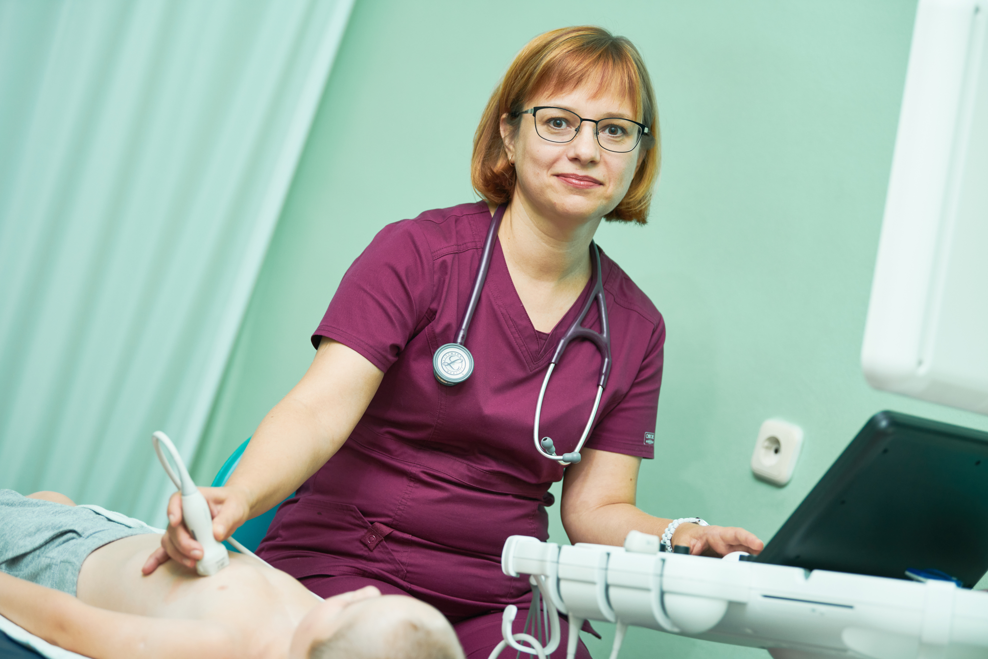
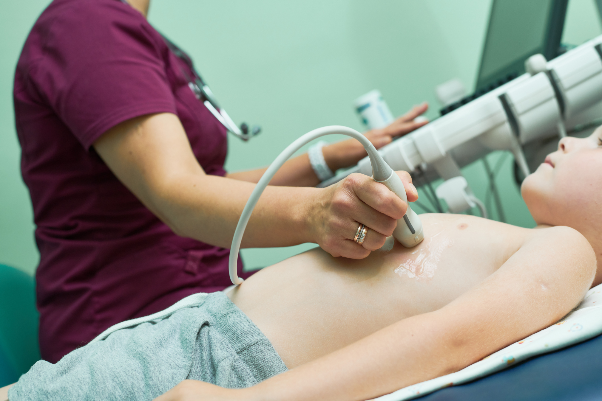
Ultrasound diagnostics room
The ultrasound diagnostics room is a structural unit of the RNPC of Pediatric Surgery and provides a wide range of modern highly informative diagnostic research methods.
Ultrasound diagnostics is a modern highly informative method of diagnosing diseases. Ultrasound methods allow for initial examination (screening), dynamic monitoring of patients, and in some cases, the results of the study are decisive in the diagnosis and choice of treatment tactics.
The ultrasound rooms are equipped with high-end and expert-class diagnostic devices. The equipment allows for all types of ultrasound examinations necessary for the examination of patients.
During the year, approximately 20,000 thousand studies are performed at the children’s center, including more than 9,000 thousand echocardiographic studies and 11,000 thousand ultrasound studies.


Staff of the ultrasound diagnostics room
Doctors from the first and highest qualification categories work in the ultrasound diagnostics room. The cabinet staff has extensive work experience. Doctors constantly take part in conferences, training, work in other clinics, etc.

Kirill Yurievich
endoscopist
(Head of the Diagnostic Department),
the highest qualification category, PhD

PAVLYUCHENYA
Olga Olegovna
doctor of ultrasound diagnostics

VARGANOVA
Regina Petrovna
doctor of ultrasound diagnostics

ZHUKOVETS
Alina Dmitrievna
doctor of ultrasound diagnostics

YUKHNEVICH
Anna Tedeushevna
doctor of ultrasound diagnostics

SNIGIREVA
Elena Vasilyevna
nurse (senior)
Range of services
Echocardiography with dopplerography and color duplex scanning (EchoCG). It is carried out in order to identify and dynamically monitor structural and hemodynamic abnormalities of the heart
Echocardiography with Doppler analysis allows you to perform an extended assessment of the work of the heart and main vessels using the following techniques:
— pulse wave Dopplerography. It is used to assess the pumping function of the heart. Allows you to record the clicks of opening and closing valves. Registers the blood flow rate in valves and main vessels;
— continuous wave Dopplerography. It allows you to measure high blood flow rates. It also serves to calculate the pressure in large vessels and cavities of the heart in different phases of the cardiac cycle;
— color dopplerography. It allows you to evaluate not only the speed, but also the direction of blood flow through the main vessels. It is used to assess abnormal blood flow through the heart valves.
Ultrasound of the abdominal cavity organs - one of the most common examination methods today is ultrasound diagnostics
With this type of examination, the doctor will be able to analyze in great detail the condition, shape, structure, size of organs and, comparing these indicators with the norm, can draw conclusions about the presence of pathology. The ultrasound protocol of the abdominal cavity includes examination of the liver, gallbladder, spleen, pancreas, kidneys and bladder.
Ultrasound of the abdominal cavity is an absolutely painless, non-invasive method of examination.
Ultrasound examination is performed on an extra-class device with a set of sensors for various organs and tissues, including sensors for the study of children aged 0 to 15 years. In newborns, the pyloric section of the stomach is evaluated for pyloric stenosis and the umbilical region for the presence of an inflammatory process.
