Computer tomography room
1st floor (office No. 2)
The Department of Radiation Diagnostics is a structural subdivision of the Russian National Center for Pediatric Surgery, is part of the diagnostic department and carries out a wide range of modern highly informative X-ray examination methods.
The X-ray examination method is based on the registration of gamma radiation in a certain spectrum (X-ray) that has passed through the human body. The use of standard radiography, even digital, does not always allow to obtain a complete picture of the pathological process due to the peculiarities of the X-ray image construction and the possibilities of the method. For example, classical radiology methods (even when working on digital media) do not allow you to create three-dimensional models. X-ray computed tomography (CT) successfully copes with these difficulties. RCT is a polypositional transmission of the body with subsequent digital processing of the received X–ray radiation that has passed through a person and an assessment of the degrees of its attenuation. During CT examination, the thickness of the slice can be up to 0.1 mm. This makes it possible to distinguish small pathological foci, including in young children.


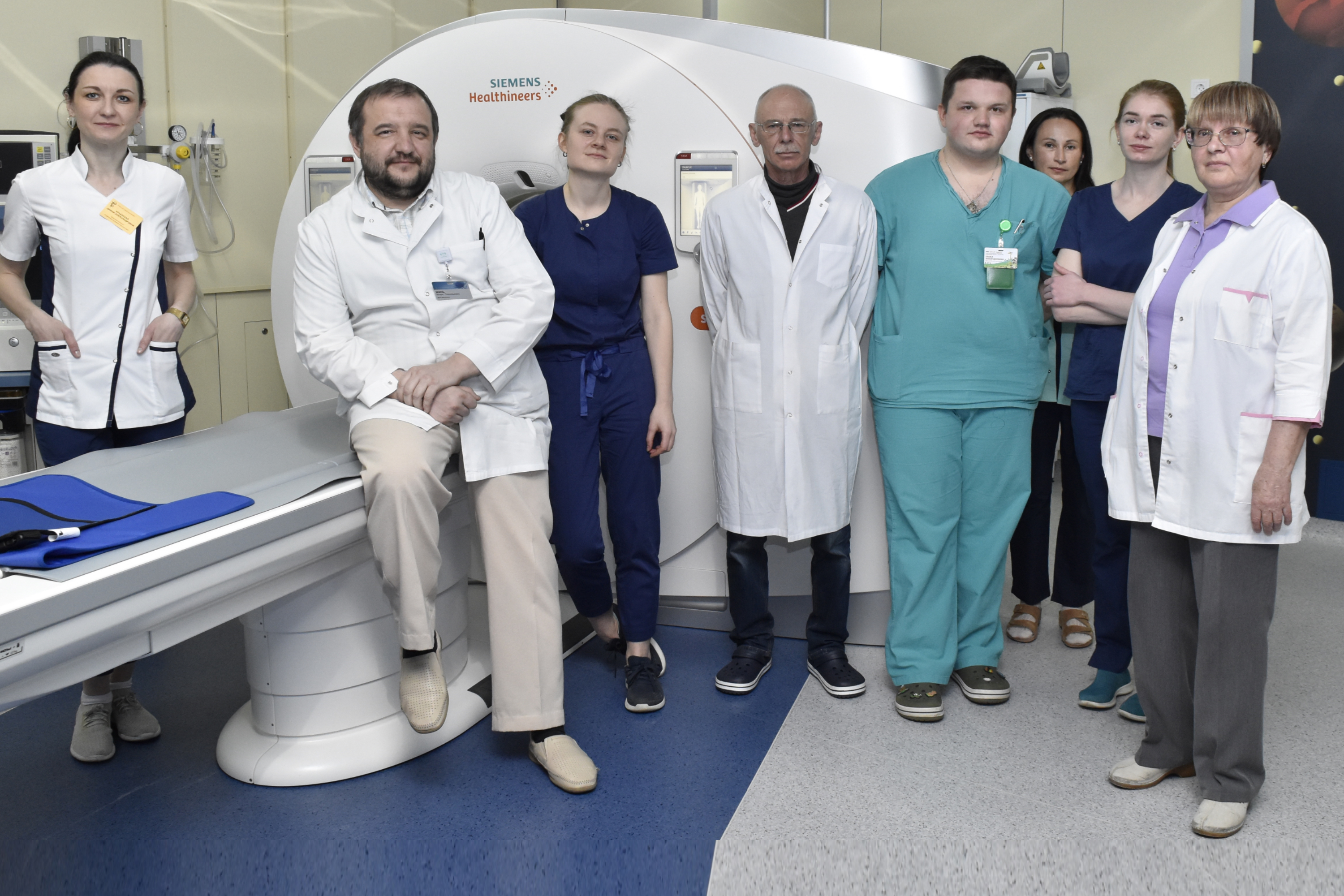
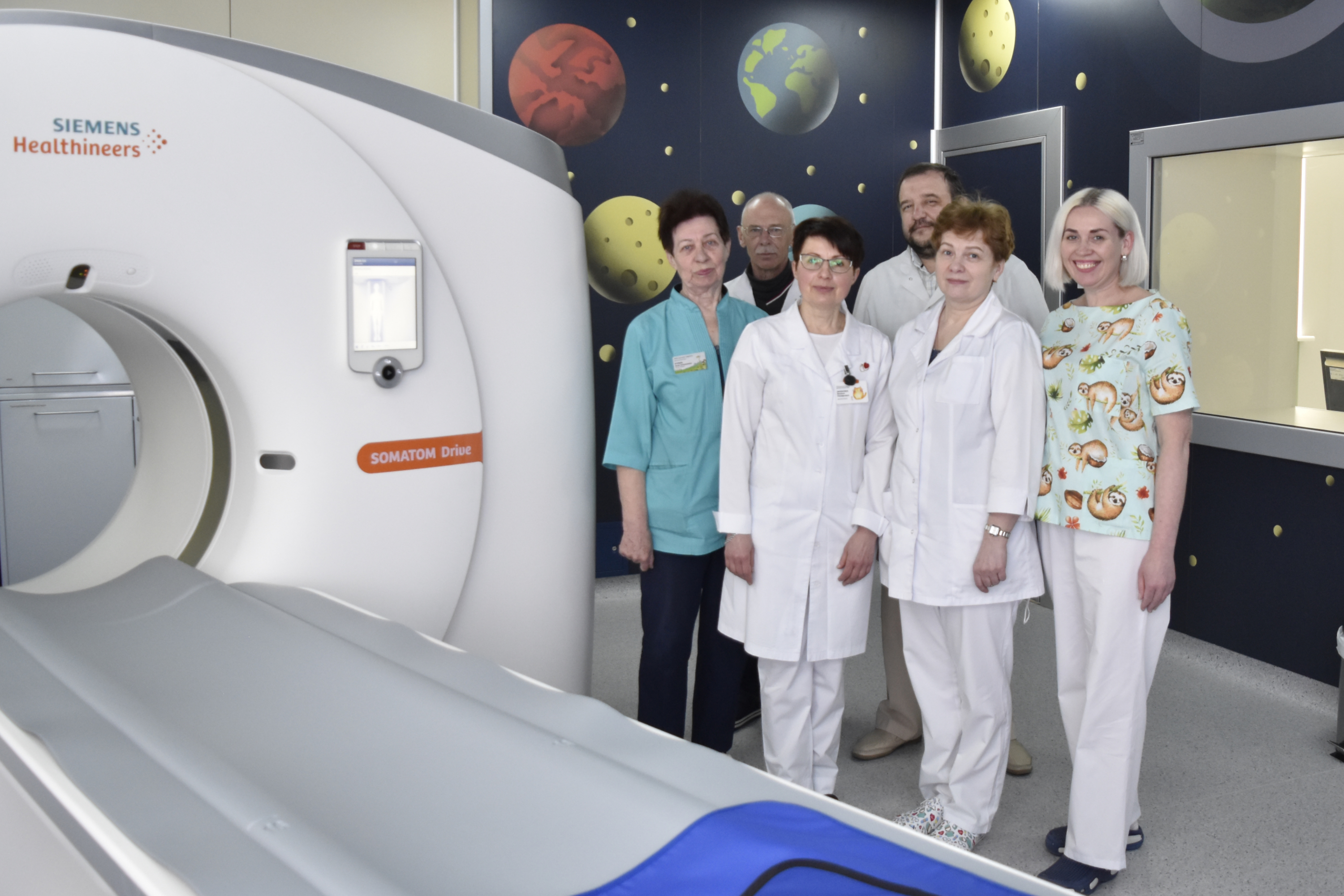
In 2022, the radiology department was equipped with modern CT machines (Siemens SOMATOM Drive). The two-tube, 128-slice, high-speed device allows you to scan in a very short period of time (a full revolution of less than half a second), almost instantly obtain high-quality images. The availability of special pediatric programs and radiation load reduction programs can significantly reduce the dose received by the patient while maintaining the proper quality of the images obtained. High scanning speed is especially important when examining the heart and blood vessels, including the vessels of the heart. High scanning speed is used to minimize artifacts (dynamic blurring). In modern devices (such as ours), cardiosynchronization is also used. RCT also allows you to assess the topography of internal organs, assess the relationship of the respiratory, digestive tract, vascular structures, the presence and degree of compression of the organs of these systems. The airways and vessels can be assessed using reconstructed multi-plane images or three-dimensional reconstructed images. It is also possible to perform dynamic computed tomography.
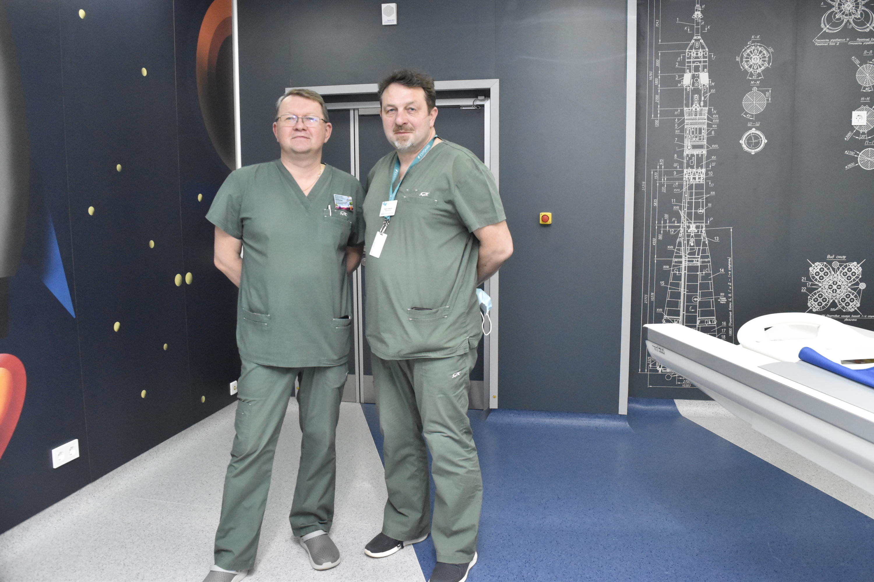
The staff of the computed tomography room
Doctors of the second, first and highest qualification categories work in the computed tomography room. The cabinet staff has extensive work experience. Doctors constantly take part in conferences, training, work in other clinics, etc.

Diana Alexandrovna
radiologist
(Head of the department)

Polina Kirillovna
radiologist

GORELCHIK
Natalia Vladimirovna
X-ray laboratory assistant (senior)
CT examination is performed by appointment (except for emergency indications), if there is a referral for research. The referral indicates the patient’s data, the rationale for the study and the presence/ absence of contraindications to the administration of contrast agents.
